Tracheostomy
Introduction
Tracheostomy is a surgically created hole through the front of the neck and into the windpipe (trachea). The term for the surgical procedure to create this opening is called as tracheotomy.
A tracheostomy provides an air passage to help the patient breathe when the usual route for breathing is somehow obstructed or impaired. A tracheostomy is often needed when some serious health problem require long-term use of a machine (ventilator) to help in breathing. In rare cases, an emergency tracheotomy has to be performed when the airway is suddenly blocked, such as after a traumatic injury to the face or neck.
When a tracheostomy is no longer needed, it can be allowed to heal shut or needs to be surgically closed. For some people, a tracheostomy is permanent.
Indications:
Medical conditions that require the use of a breathing machine (ventilator) for an extended period, usually more than one or two weeks
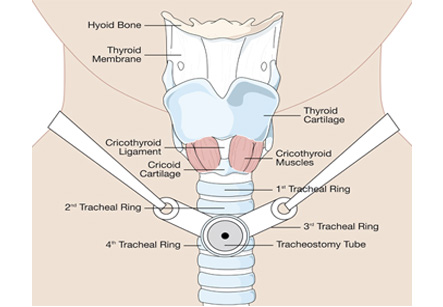
A tracheostomy is a surgical procedure to create an opening through the neck into the trachea
Conditions which block or narrow the airway, such as vocal cord paralysis or larynx cancer
Paralysis, neurological problems or other conditions which make it difficult to cough up secretions from the throat and require direct suctioning of the windpipe (trachea) to clear the airway
If patient is receiving general anesthesia, the doctor may ask to avoid eating and drinking for several hours before the procedure.
Preparation for major head or neck surgery to assist breathing during recovery
Severe trauma to the head or neck which obstructs breathing
Other emergency situations when breathing is obstructed and emergency team cannot put a breathing tube through the mouth into the trachea
Emergency care
Most tracheotomies are performed in a hospital setting. However, in the case of an emergency, it may be necessary to perform this procedure outside the hospital, such as at the scene of an accident.
Emergency tracheotomies are difficult to perform and have an increased risk of complications. A related, but somewhat less risky procedure in emergency situation is a cricothyroidotomy. This procedure creates a hole directly into the larynx at a site immediately below the Adam’s apple (thyroid cartilage).
After transfer to a hospital and stabilization, a cricothyroidostomy is replaced by a tracheostomy if there’s a need for long-term breathing assistance.
Risks:
The risks for any anesthesia are:
• Problems in breathing
• Reactions to medications
• Bleeding
• Infection
• Nerve injury
Other risks include:
• Damage to the thyroid gland
• Erosion of the trachea
• Puncture or collapse of the lung
• Scar tissue in the trachea that causes pain or breathing problems
Preparation:
Preparation for a tracheostomy depends on the type of procedure planned. If patient is receiving general anesthesia, the doctor may ask to avoid eating and drinking for several hours before the procedure. Patient may also be asked to stop certain medications.
Procedure:
A tracheotomy is most commonly performed in an operating room under general anesthesia. A local anesthetic can be sprayed to numb the neck and throat if the surgeon is worried about the airway being compromised from general anesthesia.
The type of procedure depends on the indication of tracheostomy and whether the procedure was planned or not. There are essentially two options:
Surgical tracheotomy
This can be performed in an operating room or in a hospital room. During a surgical tracheotomy, the surgeon makes a horizontal incision through the skin at the lower part of the front of the neck. The surgeon carefully retracts the surrounding muscles and cuts through a small portion of the thyroid cartilage, exposing the windpipe (trachea). At a specific location on the windpipe near the base of the neck, the surgeon creates a hole and inserts a tracheostomy tube into the hole. Faceplate and neck strap attached to the tube keep it from slipping out of the hole. Temporary sutures also can be used to secure the faceplate to the skin of the neck.
Minimally invasive tracheotomy
This is typically performed in a hospital room. The doctor makes a small incision near the base of the front of the neck. A special lens is then inserted through the mouth so that the surgeon can view the inside of the throat. Using this view of the throat, the surgeon guides a needle into the windpipe to create the tracheostomy hole. The hole is then enlarged to accommodate the tracheostomy tube. The faceplate and neck strap attached to the tube secure it from falling out of place.
Care of the tracheostomy tube
In case of permanent tracheostomy, a nurse will teach you how to clean the tracheostomy tube. In general, patient is asked to clean the tracheostomy site at least twice a day to prevent infection. This should be continued as long as there is tracheostomy tube is in place.